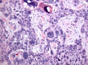
Squamous Cell Carcinoma with Monster Cells

Considering all the Monster cells, the Giant cells, the Natural Killer cells, and the Ghost cells, histopathology is scary business.  Of course I cannot identify the Natural Killer cells on H&E stained sections, I include them in my list because they sound so fierce.  If you can identify them without immunohistochemistry, you have superNatural sKills.  This biopsy shows a squamous cell carcinoma and the marked atypia and pleomorphism of the ‘monster” cells is quite interesting.  More than the usual degree of atypia anyway.  I included more high magnification shots so you can get up close with these monstrosities.  

 I admit, I missed the timing on this post and should have saved it for Halloween.

Reference

Basaloid squamous cell carcinoma with ‘monster’ cells: a mimic of pleomorphic basal cell carcinoma. Defty CL, Segen J, Carter JJ, Ahmed I, Carr RA. J Cutan Pathol. 2011 Apr;38(4):354-6.

Carcinosarcoma (Primary Metaplastic Carcinoma) of the Skin

This fascinating tumor is certainly a rare bird and, like rare birds, it seems to have multiple names: carcinosarcoma, metaplastic carcinoma, carcinoma with sarcomatous component.  Take your pick.  This lesion comes around once or twice a decade in our practice and was my first case.  There is a biphasic proliferation of malignant epithelial and mesenchymal components.  The epithelial component shows changes of superficial and solid-type basal cell carcinoma. The basal cell carcinoma component shows intimate association with a spindle cell proliferation that has a somewhat storiform pattern. The spindle cell proliferation also forms a nodule of cells that show nuclear hyperchromasia, pleomorphism and brisk mitotic activity with some atypical mitotic figures.   Immunohistochemistry studies showed no staining with epithelial markers in the sarcomatous component.

Basal cell carcinoma with a sarcomatous component (carcinosarcoma): a series of 5 cases and a review of the literature. Rose RF, Merchant W, Stables GI, Lyon CL, Platt A. J Am Acad Dermatol. 2008 Oct;59(4):627-32.

Cutaneous carcinosarcoma: adnexal vs. epidermal types define high- and low-risk tumors. Results of a meta-analysis. Tran TA, Muller S, Chaudahri PJ, Carlson JA. J Cutan Pathol. 2005 Jan;32(1):2-11

Variants

Clear cell basal cell carcinoma.  Given the never ending stream of basal cell carcinomas my eyeballs see, it is surprising to me how rarely I seen this variant of basal cell carcinoma.  The nests of tumor cells show prominent clear cell change with an adamantinoid appearance.  Usually, I will see clear cell basal cell carcinoma focally within a more typical basal cell carcinoma, but this tumor showed only clear change.  A sebaceous neoplasm may be a consideration in this case, but the cytoplasm lacks the vacuolization and nuclear scalloping of sebaceous differentiation.

Oliver GF, Winkelmann RK.  J Cutan Pathol. 1988 Dec;15(6):404-8.  Clear-cell, basal cell carcinoma: histopathological, histochemical, and electron microscopic findings.